A technology designed at Lawson Health Research Institute and Western University can unlock new doors to answer whether or not patients are reacting to treatment for enhanced ovarian cancer. A multi-centre clinical experiment has confirmed that CT perfusion that measures blood volume and blood flow to tumors linked with ovarian cancer can offer a precise calculation of how well a therapy is working, permitting doctors the chance to better plan the therapy. Capital for the experiment was offered via the National Clinical Trials Network by the U.S. National Cancer Institute counting ECOG-ACRIN and NRG Oncology.
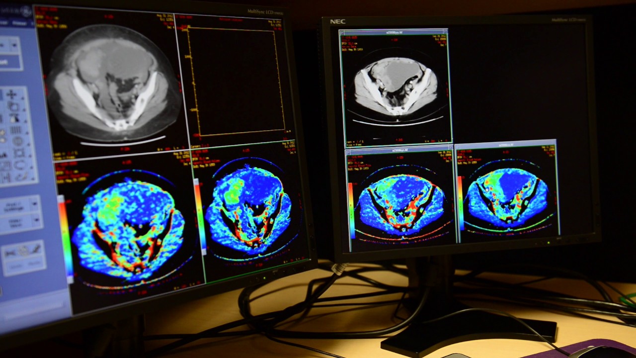
“CT perfusion is working on the change in flow of the blood to the tumor after and before the therapy,” said scientist at Lawson Health Research Institute, Ting-Yim Lee. Lee is also a medical physicist at St. Joseph’s Health Care London and a professor at Schulich School of Medicine & Dentistry of Western University. “In this specific case, we can witness that the flow of blood seems to reduce in those who will live longer without signs, whereas for those whose signs will persist within 6 months, we witnessed the flow of blood towards the tumor elevated after their therapy.”
The study was rolled out in the Clinical Cancer Research’s journal. Its authors highlighted that in 60% to 85% of patients suffering from ovarian cancer, deterioration will take place after primary therapy. Using CT perfusion to recognize which patients are more probable to profit from a specific treatment, it enables better treatment planning and selection of patient as well as offers a biomarker for upcoming clinical experiments evaluating new therapy options. The authors also highlight that even though the experiment is guaranteed, further surveys are needed to support the existing findings.
“Employing this approach, we are capable of seeing a modification in the flow of blood as early as 4 weeks post the therapy. This indicates that we do not have to wait for months to confirm whether symptoms and signs will persist, we are capable of telling whether the therapy is effectual much sooner,” claimed Lee.
Designed by Lee and his squad at Robarts Research Institute of Western University, CT perfusion employs X-ray dye in amalgamation with current CT scanners to calculate flow of the blood. The tech is already adopted all over the world to evaluate flow of the blood to the brain post the stroke. This clinical experiment for use in ovarian cancer occurred across 19 U.S. centers, and also confirmed that the tech is implemented with no trouble onto any current CT scanner, and needed least training to be victorious.